
 简体中文
简体中文 English
Website review
This review may include AI-generated content and is not individually verified. Domain, certificate and other facts come from the observed data on this page.
What is Anatomy Atelier?
Anatomy Atelier is a free, browser-based 3D human anatomy explorer built around the idea of learning anatomy "like an artist." Instead of static diagrams, it presents rotatable 3D organ models — the heart, brain, lungs, liver, kidneys, eye, intestine, pancreas and skin are among those listed — that you can manipulate directly in the page.
What you can actually do with it
- Rotate and zoom a specimen by dragging and scrolling, with an optional auto-rotate.
- Isolate a single organ or view it in cross-section and with layers.
- Click labelled points to read short explanations — for example, on the heart, dots identify the aorta, atria, ventricles and mitral valve, each with a one-line function.
- Read key facts and clinical notes for each organ: approximate size, weight, daily function, location, and common conditions such as coronary artery disease or arrhythmia.
- Compare organs side by side (the site offers a heart-versus-brain comparison) and use function animations and quizzes to test recall.
- Switch interface language, with a long list including Spanish, Hindi, Chinese, Arabic, Portuguese, French, German, Japanese, Russian, Indonesian and Korean.
Who it suits
It fits students who need a quick spatial sense of where structures sit and how they relate, art and design learners studying form, and anyone curious about basic anatomy who doesn't want to install software. The short fact panels and clinical condition lists make it a reasonable first pass before deeper textbook or lecture work. It is not positioned as a clinical, diagnostic or dissection-grade tool, so don't expect surgical-level detail or cadaver-accurate texture.
How it differs from traditional anatomy software
The site itself frames the contrast: conventional packages tend to be heavy desktop installs with dense labelling and reference-grade content, while Anatomy Atelier emphasises lightweight browser access and an exploratory, click-to-learn style. The trade-off is depth — a browser explorer is good for orientation and recall, less so for exhaustive anatomical nomenclature.
A useful next step: open the heart model, rotate it, then switch to cross-section and try naming the chambers and valves before clicking the dots. If the labels come easily, move to the quiz and the comparison view to check whether you can distinguish neighbouring structures rather than just recognise them.
How can I use Anatomy Atelier to study a specific organ like the heart or brain?
Start with the organ library, pick the heart or brain, then study it in layers: rotate and zoom the model to learn its shape and position, click the labeled dots (for the heart, structures such as the aorta, atria, ventricles and mitral valve) to read short explanations, and use isolate, cross-section and layers to see what sits inside or behind other tissue. Each organ page also gathers key facts, common conditions and a link to a fuller lesson, so you can move from "what does it look like" to "what does it do and what goes wrong."
A practical study routine
- Orient first. Rotate the whole organ and read its key facts (size, weight, location, main function) so the labels have somewhere to attach in your memory.
- Peel it back. Use cross-section and layers to move from surface anatomy inward, naming each structure as you reveal it. For the heart, trace the path of blood: right atrium, right ventricle, lungs, left atrium, left ventricle, aorta.
- Test yourself. Hide the labels or reset the view, then click each dot and try to name the structure before reading the text. The quiz and animation features support this kind of recall practice.
- Connect to the system. Open the related system view (cardiovascular for the heart, nervous for the brain) to see how the organ sits among its neighbors.
- Add clinical context last. Read the common-conditions list once you know the normal anatomy; coronary artery disease and arrhythmia mean more when you can point to the vessels and chambers involved.
Who benefits most
Artists and design students can use the rotation and cross-section tools to understand form and proportion before drawing. Students in introductory biology or anatomy courses get a free, no-install way to preview material before a lecture or lab. Curious general readers can dip into a single organ without working through a whole curriculum.
The trade-off is depth: a browser explorer with labeled points and short facts is excellent for orientation and recall, but it won't replace a textbook's detail or a dissection lab's hands-on spatial learning. If you need microscopic detail, look for the tissue views the site offers; if you need exam-level physiology, pair it with a course text.
A concrete example
Suppose you have a quiz on the heart next week. Spend ten minutes a day rotating the model and tracing blood flow from memory, then check yourself against the labeled dots. On the last two days, read the clinical notes and try to explain each condition in one sentence using the structures you've learned. That sequence—shape, then function, then pathology—mirrors how the site's own pages are organized, and it's a reasonable pattern to reuse for the brain or kidneys.
Is Anatomy Atelier free to use, and are there any paid features?
Yes. Anatomy Atelier describes itself as a free browser-based 3D anatomy explorer, and its own page includes a section titled "Is Anatomy Atelier Free?" — so the core experience of rotating, zooming, isolating and cross-sectioning organs is presented as free to use. The page evidence does not list any subscription tiers, pricing links or payment platforms, and no paid features are named anywhere in the supplied material.
What that means in practice:
- No account or download is implied. The tool runs in the browser, so a student can open it on a lab or library machine without installing anatomy software.
- The free experience centres on the organ library. Heart, brain, lungs, liver, kidneys, eye, intestine, pancreas and skin are each presented as a 3D specimen with labelled points, key facts and clinical notes.
- The learning extras are shown as part of the same product. Lessons, quizzes, function animations and organ comparisons appear alongside the 3D viewer rather than behind a separate paid tier.
- Absence of pricing signals is not proof of permanence. The page may not yet describe future premium tiers, so treat "free" as the current stated position rather than a guarantee.
A practical check before you commit
If you are choosing this for a class or a study group, open the site and confirm two things yourself: that every organ and feature you need (for example quizzes or the heart-vs-brain comparison) loads without a login prompt, and that the language selector covers your students — the page lists English, Spanish, Hindi, Chinese, Arabic, Portuguese, French, German, Japanese, Russian, Indonesian and Korean.
For a quick sense of how it sits beside other free anatomy resources, the visible organ coverage overlaps with BioDigital and InnerBody, which are worth comparing if you need a specific system or a citable reference rather than an artist-oriented explorer.
How does Anatomy Atelier compare with traditional anatomy software or textbooks for learning?
Anatomy Atelier is a free browser-based 3D explorer built for quick, visual, self-directed orientation to organs and systems, so it fits best as a first-pass learning and reference tool rather than a replacement for a structured anatomy course or a clinical textbook. Traditional anatomy software and textbooks go deeper on terminology, physiology and exam-level detail; Anatomy Atelier is strongest where a rotating model, organ isolation and clickable labels make spatial relationships click.
Where each approach fits
| Need | Anatomy Atelier | Traditional software / textbooks |
|---|---|---|
| Getting oriented fast | Rotate, zoom, isolate and cross-section organs such as the heart, brain, lungs, liver and kidneys | Often requires reading a chapter or navigating a complex interface first |
| Spatial understanding | 3D models with labeled points and comparison views | Diagrams and illustrations, usually static |
| Depth and exam detail | Key facts, clinical notes and short lessons per organ | Comprehensive physiology, pathology and terminology |
| Cost and access | Free, browser-based, no install described | Varies; textbooks and licensed software usually cost money |
| Best audience | Artists, beginners, curious learners, quick refreshers | Students in formal courses, clinicians, researchers |
A concrete scenario
An illustration student trying to place the heart's chambers and valves accurately can open the heart model, rotate it, click labeled points like the left atrium or mitral valve, and use the cross-section and layer controls to see what sits in front of what. A medical student preparing for a physiology exam will still need a textbook for pressure curves, conduction detail and pathology, but can use the model to check that their mental picture of the anatomy is correct.
Decision criterion
Choose Anatomy Atelier when the goal is spatial familiarity, visual memory or a fast refresher, and pair it with a textbook or formal course when you need mechanistic detail, clinical depth or assessment preparation. Its free, no-install browser format also makes it easy to try alongside existing study materials rather than instead of them.
As a next step, pick one organ you already know from reading and test yourself on its labeled parts in the 3D view; where you hesitate, that is the gap a textbook should fill.
For broader anatomical reference alongside it, Khan Academy and OpenStax offer free structured life-science material.
Can teachers or students use Anatomy Atelier for classroom lessons and quizzes?
Yes. Anatomy Atelier is built for exactly that kind of use: its organ pages include a View lesson option and a Quiz button alongside the 3D model, and the site frames itself around learning anatomy "like an artist." A teacher can open an organ such as the heart, rotate and isolate it on a projector, click labelled points like the mitral valve or left ventricle, and then hand the same page to students for a quiz.
Practical classroom uses
- Guided walkthroughs: Rotate, zoom and cross-section a specimen while narrating structures; the clickable dots give short definitions you can read aloud.
- Independent study: Students explore organ libraries by system (cardiovascular, nervous, respiratory, digestive, urinary and others) at their own pace.
- Quick checks: Use the quiz after a lesson, or ask students to compare two organs using the built-in comparison view.
- Art and biology crossover: The "learn like an artist" framing suits drawing classes studying form and proportion as much as biology revision.
Trade-offs to weigh
The tool is strongest for surface-level structure and vocabulary — organ identification, key facts, common conditions. It is not a substitute for dissection, histology slides or detailed physiology teaching. The excerpt shows clinical notes and a microscopic tissue view, but you should confirm depth against your syllabus before making it a primary resource. Browser-based access also means you depend on working devices and connectivity in the room.
A sensible next step
Pick one lesson you already teach — say, the heart — and preview its 3D page, lesson and quiz yourself first. If the quiz questions match your learning objectives, assign it as pre-class preparation so class time goes to discussion and drawing. For broader anatomy references, compare with InnerBody or Visible Body if you need more detailed or paid institutional content.
What interactive 3D features does Anatomy Atelier offer, such as cross-sections, layers, or comparisons?
Anatomy Atelier centres its 3D explorer on a set of manipulation tools rather than a single rotating model. According to the site, the viewer supports rotate, zoom, isolate, cross-section, layers, compare and reset, with an auto-rotate option and clickable anatomy dots that open labelled notes.
What each feature is for
- Rotate and zoom: drag to rotate and scroll to zoom, so you can inspect an organ from several angles or move in on a region. The page presents this as the basic way to study a specimen.
- Isolate: show one structure on its own. This suits a learner trying to fix the shape and position of a single organ before adding surrounding anatomy.
- Cross-section: look inside a model rather than only at its surface. The heart example implies cutting through chambers and valves, which is more useful for internal structure than a surface view.
- Layers: peel back or add anatomical levels. This is the feature to check first if you need to move from skin or muscle down to deeper structures.
- Compare: the site shows an "Open comparison" entry and a "Heart vs. brain" example, suggesting side-by-side study of two organs. It is aimed at contrast — for instance, comparing how two organs are supplied with blood or organised internally.
Clickable labels and supporting material
The heart model includes dots for the aorta, left and right atria, ventricles and mitral valve, each with a short explanation. Each organ page also carries key facts (size, weight, location, function), clinical notes on common conditions, a tissue view such as cardiac muscle, and links to an animated function and a quiz. The organ library spans the heart, brain, lungs, liver, kidneys, eye, intestine, pancreas and skin, grouped by body system.
Who benefits, and the trade-off
A student revising for a biology or anatomy exam gets the most from isolate, cross-section and quiz together: isolate a structure, cut through it, then test recall. An artist or illustrator will lean on rotate, zoom and layers to judge form and depth. The limitation is that a browser model is a study aid, not a substitute for dissection, histology slides or clinical imaging; the clickable dots give concise facts rather than deep detail.
As a next step, open the heart model, isolate it, switch on the cross-section and click each labelled dot in turn, then use the comparison view against the brain to see how differently the two organs are structured.
 Cloudflare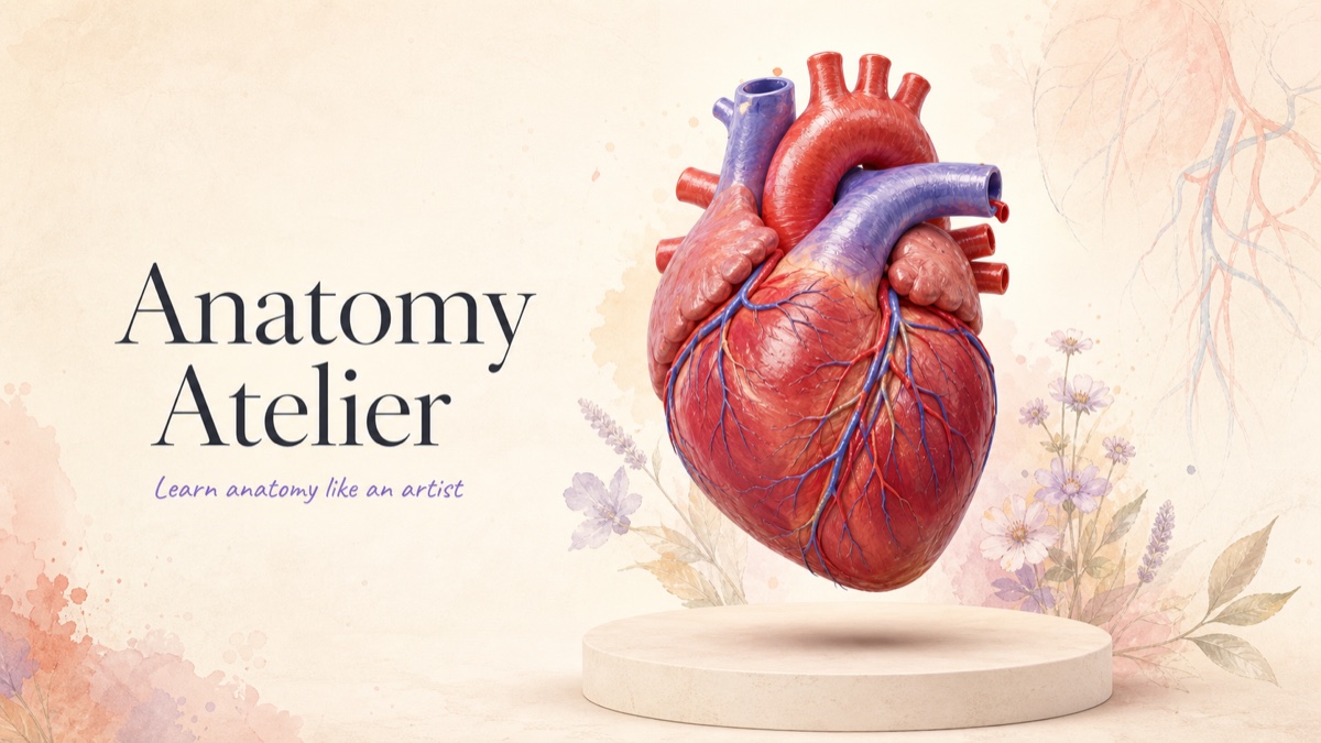

User reviews (0)